Understanding Calcium Deposits in the Shoulder and Their Implications
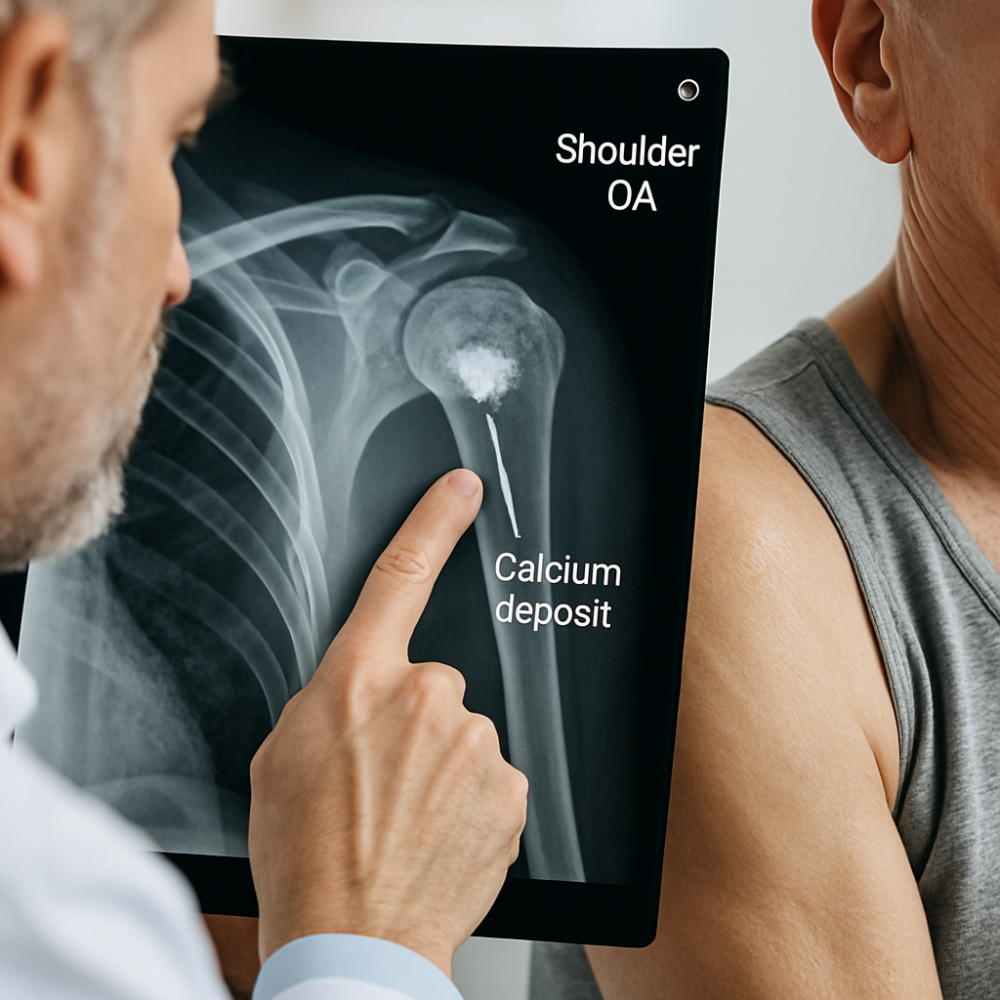
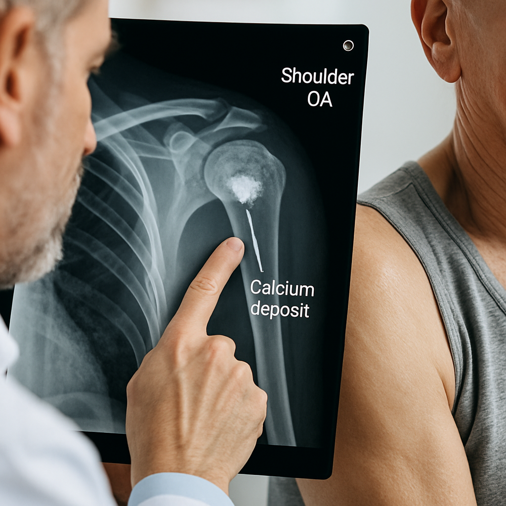
Shoulder calcium deposit removal is a minimally invasive procedure designed to target and eliminate calcified buildups that develop within the rotator cuff tendons, most commonly the supraspinatus. These deposits, which can be visualized on an x-ray and are often associated with pain or dysfunction, may be linked to conditions such as osteoarthritis or degenerative rotator cuff changes. For patients suffering from persistent joint pain and loss of range of motion, surgical removal offers effective symptom relief and helps restore functional abilities. The underlying mechanism involves removing not only the calcium but also addressing coinciding tendon or joint pathology if needed, supporting a faster return to normal activity. For a detailed patient account and further explanation of the procedure, refer to the Sydney Shoulder Unit.
Diagnosing Causes of Deposits and Imaging with X-Ray
Calcium deposits within the shoulder can result from a range of metabolic and mechanical factors. Common contributors include chronic microtrauma to the tendon, reduced blood supply in specific regions of the tendon, or underlying osteoarthritis, which creates an environment conducive to the precipitation of minerals like calcium phosphate. Understanding these causes is crucial to determining whether medical management or a surgical solution is warranted.
X-ray imaging remains the primary modality for identifying calcified deposits in rotator cuff tendons. On radiographs, these deposits present as discrete, high-density areas. Sometimes, further imaging using MRI or ultrasound can clarify the size, location, and degree of associated inflammation or tendon involvement, providing essential information for surgical planning. According to orthopedic specialists, such as featured on the Dr. Meyer Orthopaedics site, selecting the right imaging protocol can enhance precision and outcomes in treatment.
It is important to distinguish calcific tendinitis from other shoulder conditions like costochondritis or rheumatoid arthritis, as management strategies and prognosis may differ. Combining clinical evaluation with advanced radiology allows clinicians to tailor interventions effectively and avoid unnecessary procedures.
Patients may also undergo serial x-rays or other imaging techniques postoperatively, especially if symptoms persist or there is concern for incomplete removal or recurrence. Monitoring for additional joint changes remains essential for long-term health.
Minimally Invasive Approaches for Rotator Cuff Calcium Shaver Treatment
Arthroscopic techniques are the gold standard for shoulder calcium deposit removal. This approach utilizes a small camera and specialized tools, such as an arthroscopic shaver, to precisely excise the calcific material while minimizing trauma to healthy tissue. By operating through tiny incisions, surgeons reduce scarring risk, postoperative pain, and facilitate rapid recovery as outlined on the Sydney Shoulder Unit page.
During the operation, the tendon is carefully probed. If there are concurrent issues, such as tendon tears, biceps involvement, or the presence of osteoarthritis seen on imaging, these can be addressed simultaneously without the need for additional surgeries. This comprehensive approach helps ensure the shoulder is functionally optimized before the patient begins rehabilitation.
For those with extensive calcification, especially when combined with rotator cuff degeneration or metabolic bone disorders like osteoporosis, a tailored approach might include adjunct therapies or implantable materials (such as scaffold grafts or bone substitute supplements) to aid tendon healing.
The Recovery Process: From Sling to Full Range of Motion
Immediate postoperative management typically involves protecting the shoulder using a sling. Most patients can expect to use this for up to two weeks, during which time sutures and dressings keep the small keyhole incisions safe. However, early initiation of gentle, assisted range of motion is encouraged, following expert physiotherapy guidelines like the JPL protocol. This enhances healing and helps prevent joint stiffness and tetanic contraction.
At the two-week mark, formal physiotherapy is commenced, focusing on progressive increases in shoulder movement. Most patients are able to regain functional range within weeks, particularly if there is no extensive tendon damage or comorbid osteoarthritis. Pain management usually does not require long-term medications, and rapid weaning from the sling is often possible.
Complications are uncommon but may include postoperative stiffness or, rarely, nerve injury. According to Dr. Meyer Orthopaedics, most cases resolve with patience and appropriate therapeutic interventions. Persistent symptoms or recurrent deposits may need further evaluation, sometimes necessitating repeat x-ray.
Metabolic Factors and Osteoarthritis in Calcium Formation
Several systemic factors contribute to the formation of calcium deposits in the shoulder. Chronic conditions like osteoporosis or disturbances in mineral metabolism can increase susceptibility, as can dietary patterns low in essential bone and joint supplements. Additionally, bisphosphonates (often prescribed for bone health) have complex interactions with both bone and tendon tissue and require careful consideration if you are undergoing or recovering from this type of surgery.
Addressing these root causes is essential not only for preventing recurrence but also for optimizing overall musculoskeletal health. Patients may benefit from nutritional interventions, physiotherapy tailored to their unique biomechanics and posture, and ongoing evaluation of comorbid conditions such as osteoarthritis or even rare causes like parathyroid dysfunction. Serial x-ray follow-ups can help track any new or residual deposits and inform appropriate management tweaks.
Shoulder Health Maintenance: Prevention of Recurrence and Long-Term Outcomes
Successful calcium removal combined with physical rehabilitation and lifestyle modification typically restores near-normal function for most patients. Preventative strategies include maintaining optimal shoulder posture, participating in periodic physiotherapy, and managing systemic diseases (such as osteoporosis or rheumatoid arthritis) that elevate the risk of further calcification or joint pain deposits.
Clinicians may recommend regular supplement intake, use of instrumental therapies for muscle balance, and periodic imaging to catch any early signs of recurrence. Long-term results are generally positive, with many patients returning fully to sport and daily life, though those with severe underlying osteoarthritis or metabolic disturbances may require more frequent monitoring. Overall, prompt and effective surgical care, assisted by advanced radiology and comprehensive aftercare, provides the best outcomes in managing shoulder calcium deposits.
For more details and tailored advice, be sure to check the expert resources at Sydney Shoulder Unit.
One thought on “What to Expect in Shoulder Calcium Deposit Removal: Osteoarthritis, X-Ray & Rotator Cuff Insights”