Understanding Labeled Diagrams of the Digestive System
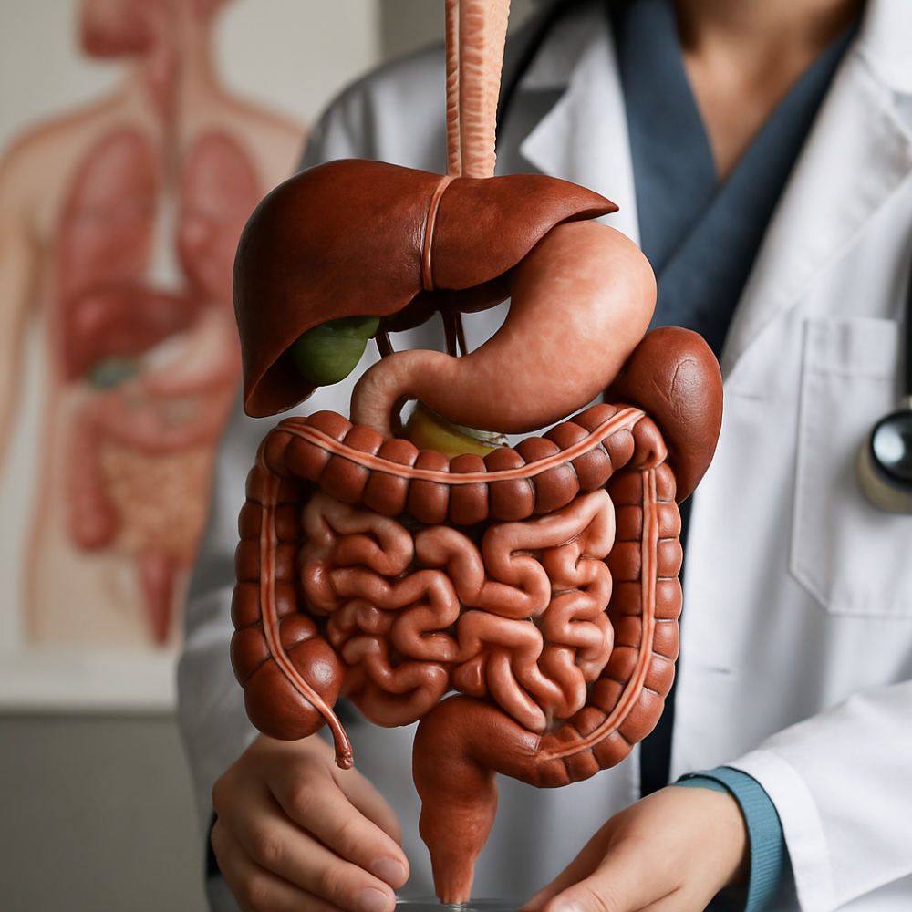
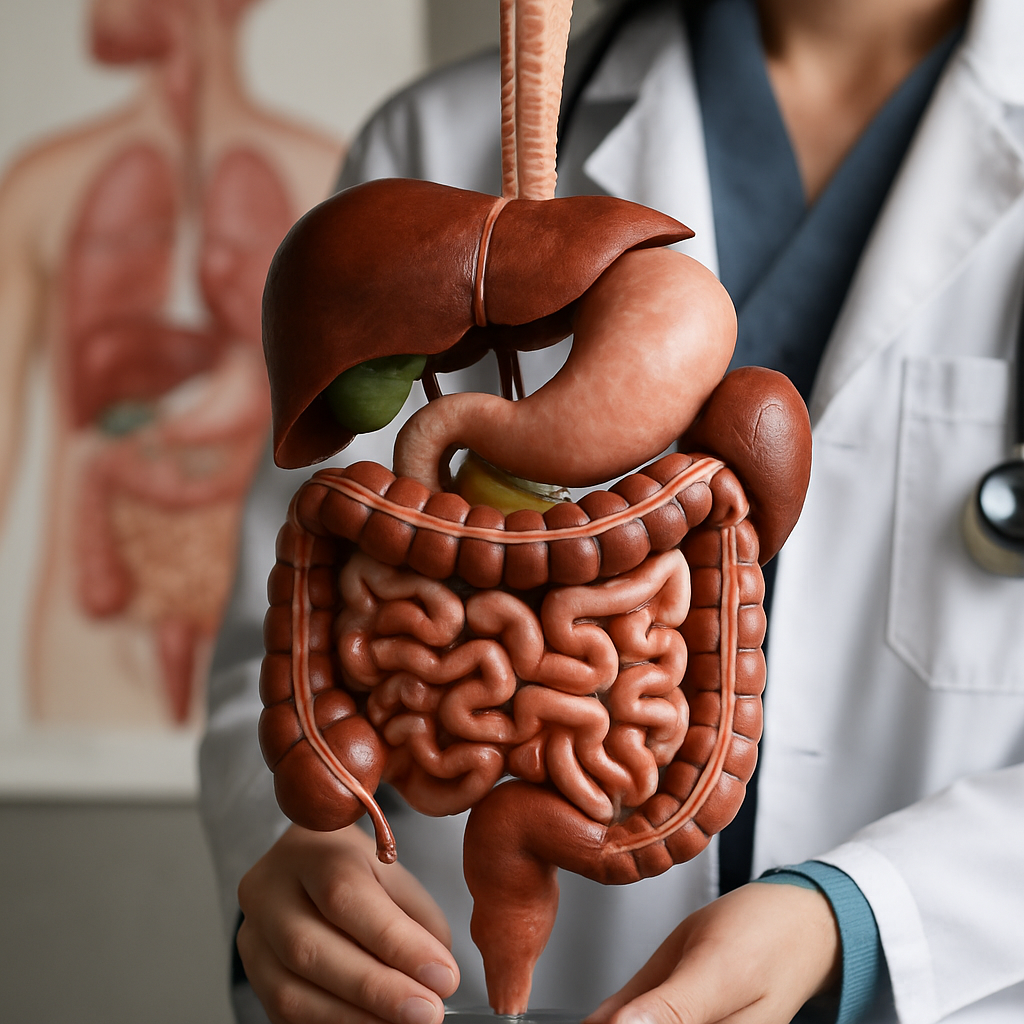
If you are looking for pictures of the digestive system with clearly labeled parts, you will find that accurate diagrams provide invaluable insight into how the body’s organs work together. These illustrations often prominently display key sections such as the mouth, esophagus, liver, stomach, gallbladder, pancreas, small intestine, large intestine, rectum, and anus. Having a visual reference makes it easier for students, health professionals, and anyone interested in human biology to understand the complex digestive process from start to finish.
Digestive system images, like those found in reputable government health resources, show how each part connects and functions. For those who want a trustworthy source, the National Institute of Diabetes and Digestive and Kidney Diseases provides a clear, labeled diagram that highlights all critical organs. Such visual resources also serve as a foundation for learning about digestive tract health, diseases, and treatment strategies.
In addition to anatomical accuracy, these diagrams often identify both primary digestive organs and accessory organs such as the pancreas and gallbladder, helping users see not just where each organ is, but also how they cooperate to break down food, absorb nutrients, and expel waste efficiently.
Key Organs in Digestive System Diagrams
Every thorough digestive system illustration will label the mouth as the entry point for food and the start of mechanical and chemical digestion. The esophagus connects to the stomach, serving as the conduit for swallowed food. The liver and gallbladder, situated near the stomach, are crucial for bile production and storage, underscoring their role in fat digestion and metabolic processing.
The stomach itself is depicted as a muscular organ that churns and mixes food with gastric acids, starting protein digestion. Diagrams also show the pancreas positioned behind the stomach, where it secretes digestive enzymes that act on proteins, fats, and carbohydrates. This helps manage not just digestion, but also blood sugar through hormonal regulation. You can find comprehensive anatomical notes about these organs in the American Cancer Society gallery.
The small intestine, typically illustrated as the longest part of the system, is responsible for nutrient absorption. Its three segments—the duodenum, jejunum, and ileum—work in a sequence to absorb water and nutrients before food remnants pass into the large intestine for further water absorption and eventual elimination as waste.
Gallbladder Attack and Digestive Enzymes in Context
During digestion, the gallbladder releases bile—in response to fatty foods—into the small intestine, which aids in digesting fats. Diagrams that highlight the gallbladder can help users visualize its location under the liver and its connection via the bile ducts. The interplay between the liver, gallbladder, and small intestine is essential knowledge for understanding conditions like gallbladder attacks, which can cause pain if gallstones obstruct bile flow.
In addition to bile, digestive enzymes produced by the pancreas break down proteins, fats, and carbohydrates. Labeled diagrams typically show the pancreas and its ductwork feeding into the duodenum. This is where most over-the-counter enzymes act—supplementing your body’s natural ability to handle complex foods, especially for individuals with pancreatic insufficiency or digestive issues such as bloating and food-related discomfort.
Interesting Facts Visible in Digestive System Pictures
Many digestive system images offer more than just anatomical labels; they sometimes include cool facts or highlight unique features like the length of the small intestine or variations in acid production across individuals. For example, fun details may include how the large intestine reclaims water from digested material, and how the rectum and anus are the final points of waste expulsion in the digestive tract.
Some diagrams emphasize key clues for identifying and diagnosing digestive diseases using labeled organ pathways, underlining the importance of comprehensive imagery in medical education and patient care. Such annotated visuals can also help explain why certain symptoms—for example, abdominal pain from a gallbladder attack—are felt in particular body areas.
By examining trustworthy and detailed images, you become better equipped to understand a range of digestive process nuances, helping you connect symptoms, such as chest discomfort or acid reflux, to underlying digestive mechanisms. This is particularly important for those exploring the causes and solutions to conditions like constipation or abdominal discomfort.
How to Use Digestive System Pictures for Learning
Learning from labeled images enhances the retention of key digestive concepts by linking anatomical structure to function. Students and educators benefit from referring to high-quality sources and images, which reinforce lessons about where enzymes act, how bile is released, and how nutrients move through the body. Such diagrams simplify complex topics, making self-study or formal education more accessible and engaging.
For healthcare professionals, consistently referencing anatomical illustrations ensures accuracy when educating patients about their digestive health, treatment plans, or surgical interventions involving organs like the gallbladder or pancreas. Visuals also play a role in framing dietary advice, especially about foods that trigger issues like acid reflux or bloating.
Whether for academic, clinical, or personal learning, authoritative resources like the NIDDK digestive system media asset or anatomical galleries provide the most reliable foundation for understanding and visualizing the full digestive tract.
Ultimately, pictures of the digestive system—with labels highlighting organs, pathways, and connections—offer an essential tool for decoding how food moves from ingestion to digestion, nutrient absorption, and eventual waste elimination.